Cancer Technologies
T Cell Biology for Cancer
T cell mechanomemory for cellular immunotherapy
Thursday, October 9, 2025
2:30 PM - 2:45 PM PDT
Location: Room 24B

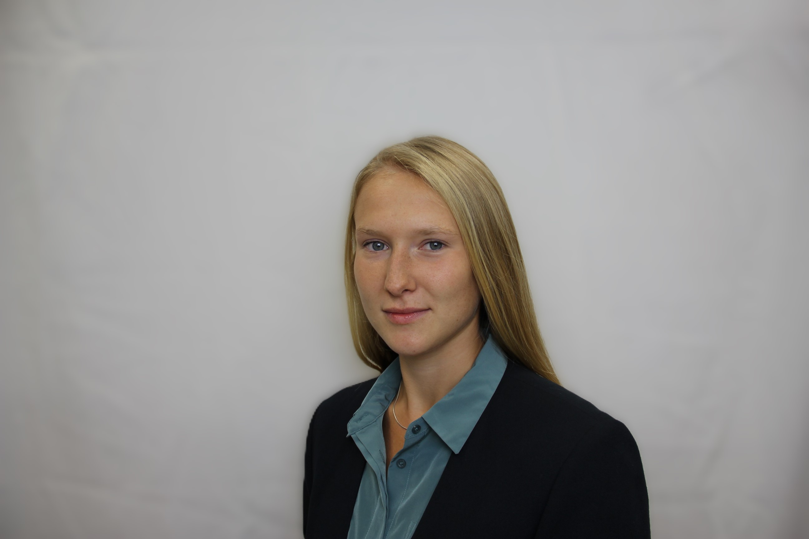
Anna-Liisa Sepp
Graduate Student
Columbia University
New York, New York, United States
Lance C. Kam
Professor
Columbia University
New York, New York, United States
Presenting Author(s)
Primary Investigator(s)
Introduction: : T cells are highly successful agents for clinical applications involving targeted immune-mediated cytotoxicity. However, a persistent challenge in adoptive cell therapy is overcoming the barriers presented by solid tumors. Notably, the mechanical properties of the tumor microenvironment (TME) are known to impair T cell activation and proliferation [1]. Here, we aim to investigate how T cell mechanosensing can be leveraged to enhance therapeutic function, with a focus on modulating ex vivo activation methods. Previous data from our group suggests that T cells retain a form of "mechanical memory", where cells retain a lasting imprint of the mechanical activation conditions that continues to influence their proliferation for multiple cell divisions after removal from the activating substrate [2], [3]. Here, we explore a hypothesis that T cells extrinsically modify their environment via stiffness-dependent media conditioning, resulting in a form of media-driven mechanomemory. We look to demonstrate how T cells condition their media environment as a result of mechanically-tuned activation as a paracrine vehicle for instilling mechanomemory. These mechanisms have important implications for optimizing T cell expansion protocols in the context of adoptive immunotherapy.
Materials and
Methods: : Mechanomemory was evaluated by proliferation and cell function following activation on engineered mechanically-tuned substrates. Human CD3+ T cells were activated on polydimethylsiloxane (PDMS) substrates of two stiffnesses: soft (1:50 PDMS, 65kPa) and stiff (1:10 PDMS, 4MPa), coated with anti-CD3/anti-CD28 antibodies. After three days of activation, cells were transferred to standard tissue culture plates and diluted every two days.
In media-switch experiments, cells were cultured in conditioned media collected from T cells previously activated on the opposite stiffness substrate. Conditioned media was harvested on day 3 or 5 by centrifugation to remove cells, then used to resuspend the target T cell population. For restimulation studies, cells were expanded as above until day 9, when cells were determined to return to quiescence (below 10fL volume), then normalized in concentration and restimulated on either matched or mismatched stiffness substrates. Proliferation was quantified using hemocytometer cell counts, while differentiation status was evaluated by flow cytometry for CD45RA, CD62L, CD4, and CD8 expression. Cytokine secretion was analyzed using short-term capture assays or multiplexed Luminex ELISA of culture supernatants at defined time points.
Results, Conclusions, and Discussions:: Our findings show that T cell mechanomemory is governed by a combination of intrinsic and extrinsic factors. T cells activated on soft substrates (65 kPa PDMS) achieved higher cumulative doublings than those on stiff substrates (4 MPa PDMS) across most donors (n = 10; 6 of 10 donors p < 0.05). Whereas stiff-activated T cells typically peaked in expansion by day 9, soft-activated cells continued expanding through day 13, indicating that early mechanical cues remain imprinted in T cells well beyond the initial activation window.
To investigate the contribution of extrinsic factors, we conducted media-switching experiments during early expansion. T cells initially activated on stiff substrates showed rescued proliferation when switched into soft-conditioned media; conversely, soft-activated cells exhibited reduced expansion when switched into stiff-conditioned media (n = 8 donors; 6 donors showed significant modulation, p < 0.05). Switching media on days 1 or 3 significantly impacted expansion outcomes, while switching at day 5 had lessened effect, demonstrating a critical time window during which mechanomemory is determined during early activation (Fig. 1). We further examine how media switching influences T cell differentiation (e.g., CD45RA expression, Tnaive:Teffector:Tmemory ratios) and functional outputs (e.g., killing capacity, exhaustion markers) at late timepoints as downstream consequences of mechanomemory. Preliminary findings reveal longer maintenance of naive markers (CD45RA) in soft-activated T cells as well as T cells transferred to soft-conditioned media, suggesting that soft and stiff substrates can influence cell differentiation via media conditioning (Fig. 2). We also present ongoing work that evaluates the identity of secreted soluble factors in conditioned media and how mechanical activation substrates drive variability in cytokine profiles.
In parallel, we explore an intrinsic, cell-internal component of mechanomemory. Following nine days of expansion on defined stiffness substrates, T cells restimulated on matched or mismatched substrates retained proliferation reflective of their initial activation condition, supporting the existence of a long-term mechanical imprint (n=2 donors).
T cell mechanomemory has significant translational relevance in cell therapy manufacturing. Leveraging innate mechanisms of mechanical memory demonstrated here can enable improved ex vivo T cell production and attack of solid tumors in patients receiving cell therapy.
Acknowledgements and/or References (Optional):: [1] J. Rojas-Quintero et al., “Car T Cells in Solid Tumors: Overcoming Obstacles,” Int. J. Mol. Sci., vol. 25, no. 8, p. 4170, Apr. 2024, doi: 10.3390/ijms25084170.
[2] L. Shi, J. Y. Lim, and L. C. Kam, “Substrate stiffness enhances human regulatory T cell induction and metabolism,” Biomaterials, vol. 292, p. 121928, Jan. 2023, doi: 10.1016/j.biomaterials.2022.121928.
[3] D. J. Yuan, L. Shi, and L. C. Kam, “Biphasic response of T cell activation to substrate stiffness,” Biomaterials, vol. 273, p. 120797, Jun. 2021, doi: 10.1016/j.biomaterials.2021.120797.
