Tissue Engineering
Biomaterials in Tissue Engineering I
A 3D Bioprinted Glioblastoma Model with Tunable Hypoxic Microenvironment
Thursday, October 9, 2025
1:30 PM - 1:45 PM PDT
Location: Room 29A

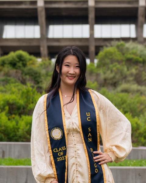
Emmie J. Yao
Graduate Researcher
UC San Diego, United States- SC
Shaochen Chen, Ph.D.
UC San Diego, United States
Presenting Author(s)
Primary Investigator(s)
Introduction: : Glioblastoma Multiforme (GBM) is the most common malignant brain and central nervous system tumor with grim prognosis and high recurrence rates. Its dynamic ecosystem contains high intratumor and interpatient heterogeneity, consisting of hallmarks such as hypoxia and necrosis that often strengthen the tumor. With this, there is an urgent need to develop GBM models that are physiologically and clinically relevant and use them to uncover underlying driving mechanisms of the disease and develop patient models to tailor treatment plans. In the past few decades, GBM organoids have emerged as a model to increase cellular heterogeneity compared to traditional 2D cultures, however, they are often low throughput and lack reproducibility. Here, we harnessed rapid 3D bioprinting to create biomimetic GBM models that recapitulate the mechanical and structural properties of native tumor microenvironment. By manipulating cell density, we were able to build off this base GBM model and create high cell density (HCD) constructs to include a key GBM hallmark - pathological hypoxia - which arises due to lack of oxygen and nutrients. Paired with chemotherapy and radiotherapy testing, our approach offers a reproducible method to model hypoxia-driven GBM progression and lays the groundwork for future therapeutic testing.
Materials and
Methods: : Human patient derived glioblastoma stem cell CW468 and human GBM immortalized U87 cells were individually encapsulated within 5% GelMA (95% degree of methacrylation) bioink with LAP photoinitiator to achieve a printable hydrogel formulation. Bioprinting was performed using a digital micromirror device system to generate 3D constructs with high spatial resolution and high throughput. Constructs were cultured under standard conditions and analyzed at defined time points. Gene expression was evaluated via qPCR using primers including HIF1a, VEGFA, CXCL12, and other GBM-specific markers. Invasion assays were conducted to quantify the migratory capacity of CW468 cells, using GFP to visually track their movement throughout one week. To assess chemotherapeutic response, bioprinted models were treated with temozolomide (TMZ) and cell viability was measured using CellTiter-Glo assay, with luminescence quantified via a TECAN plate reader. All experiments were performed in biological triplicates unless otherwise specified.
Results, Conclusions, and Discussions:: Mechanical characterization of our GBM models demonstrated that with our tailored bioink formulation, combined with optimized exposure time and cell density, yields physiologically relevant stiffnesses that closely mimic native GBM tissue. Building upon this base model, we developed an HCD model with GSCs that pathologically establishes a hypoxic microenvironment over several days of culture, as shown by the upregulation of HIF1α expression measured by qPCR. The U87 cell line did not show an upregulation of HIF1α, indicating that either a greater cell density is required to form this hypoxic microenvironment, or U87 cells are more robust in nature. Due to the overall goal of using patient-derived GBM cells, we decided to proceed with only the GSC line CW468. Invasion assays revealed that the HCD group exhibited significantly greater invasiveness, with a 40% invasion area extending beyond the original bioprinted construct, compared to just 3% in the base model. These results suggest that the HCD environment may contribute to a more aggressive tumor phenotype. Drug response studies using the standard-of-care chemotherapeutic agent TMZ showed that both the base and HCD models exhibited increased resistance compared to traditional 2D cultures, indicating that these GBM cells are more resilient in the 3D microenvironment. We are currently expanding our investigation by testing additional GBM-targeted drugs, incorporating radiotherapy to further evaluate therapeutic responses within our models, and adding additional GBM cell types.
Ultimately, our goal is to establish a platform for personalized treatment testing by integrating patient-derived GBM cells into our 3D bioprinted constructs. This study demonstrates the potential of 3D bioprinting to generate GBM models that replicate both the mechanical and pathological features of native tumors, offering a robust tool for studying tumor progression and therapeutic resistance.
Acknowledgements and/or References (Optional)::
